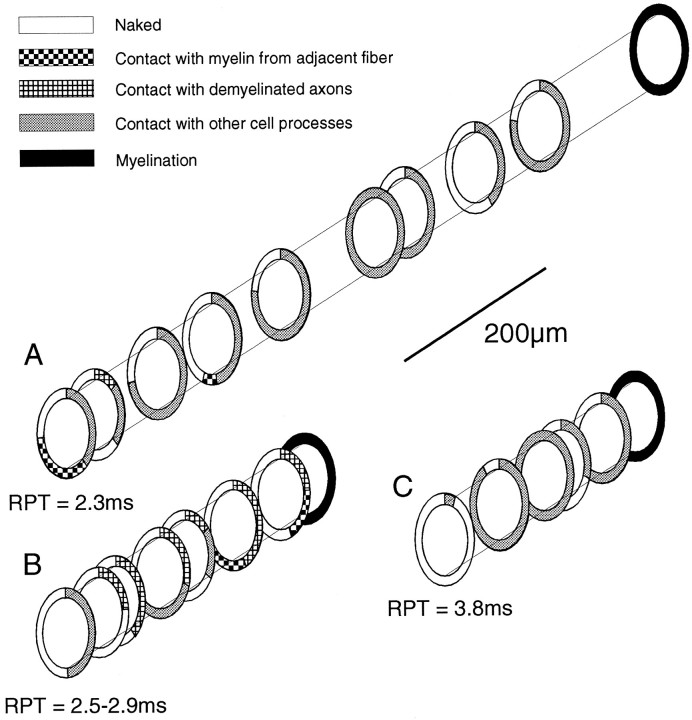
Fig. 7.
Diagram illustrating the type of ensheathment surrounding the labeled portion of three axons with known RPTs; the RPTs through the lesion are shown below each axon. The type of ensheathment was measured from electron micrographs; theNaked category includes axons surrounded by vesicular myelin debris (e.g., Fig. 5F). Lesion ages wereA, 21 d; B, 19 d; andC, 28 d.

