
Fig. 2.
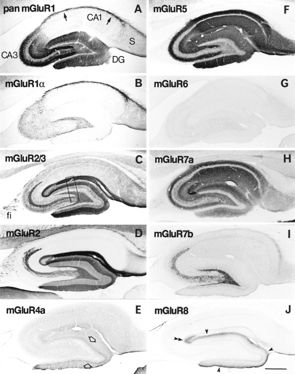
Distribution of immunoreactivity for eight mGluRs in rat hippocampus. Parasagittal sections through the hippocampus were reacted with antibody to pan mGluR1 (A), mGluR1α (B), mGluR2/3 (C), mGluR2 (D), mGluR4a (E), mGluR5 (F), mGluR6 (G), mGluR7a (H), mGluR7b (I), or mGluR8 (J). Immunoreactivity for mGluR5 (F) and mGluR7a (H) is distributed in all dendritic layers throughout the hippocampus, whereas immunoreactivity for the other mGluRs is restricted to distinct regions. Immunoreactivity for mGluR1 (A) is strong in dendritic fields of the dentate gyrus (DG) and CA3, as well as in the CA1 stratum oriens/alveus border (arrows). Immunoreactivity for mGluR1α (B) is strong only in the CA1 stratum oriens/alveus border (see Results). Immunoreactivity for mGluR2/3 (C) and mGluR2 (D) is strong in terminal zones of the perforant path and mossy fibers (see Results), whereas that for mGluR7b (I) is restricted to the mossy fiber terminal zone, and that for mGluR8 (J) to the lateral perforant path terminal zone, i.e., the outer third (filled arrowheads) of the dentate molecular layer and outer layer (double arrowhead) of CA3 stratum lacunosum moleculare. Immunoreactivity for mGluR4a (E) is weak but prominent in the inner third (open arrows) of the molecular layer. Labeling for mGluR6 (G) is hardly detected in the hippocampus. The rectangle inC indicates a corresponding region shown with a higher magnification in Figure 3. fi, Fimbria;S, subiculum. Scale bar, 500 μm.