
Fig. 2.
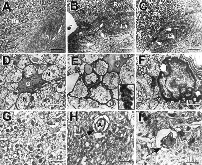
Multilayered glial membranes in swsmutant brains as seen in EM horizontal sections. A, D, G, Wild type. A, Retina and lamina.D, Cell bodies of the lamina cortex, showing a glial cell recognizable by its dark cytoplasm. Extensions of the glial cell surround various neurons with single-layered wrappings.G, Medulla neuropil, a tangle of synapses. B, E, H, In newly eclosed sws1flies, glial processes form multiple sheaths around neurons in the cortex (arrows in B, inset inE) and around axons in the neuropil (arrow inH). C–I, In 7-d-oldsws1 flies, large membrane whorls are visible (F, I). These are especially prominent in the lamina cortex (arrows in C). Flies were kept at 25°C. Re, Retina, La, lamina; G, glial cell; N, neuron;A, axon. Scale bars: A–C, 3 μm;D–I, 0.5 μm.