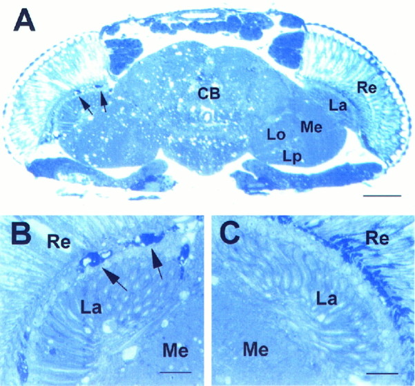
Fig. 4.
Head section ofsws4 mosaic fly, 8 d old.A, Left side, B, Darkly stained bodies characteristic of the sws mutation (arrows). These are missing toward the wild-type side at the right (A, C). The few vacuoles on the wild-type side may be caused by mutant neurons projecting from the mutant side to the wild-type side.Re, Retina; La, lamina;Me, medulla; CB, central brain;Lo, lobula; Lp, lobula plate. Scale bars:A, 50 μm; B, C, 10 μm.

