Fig. 6.

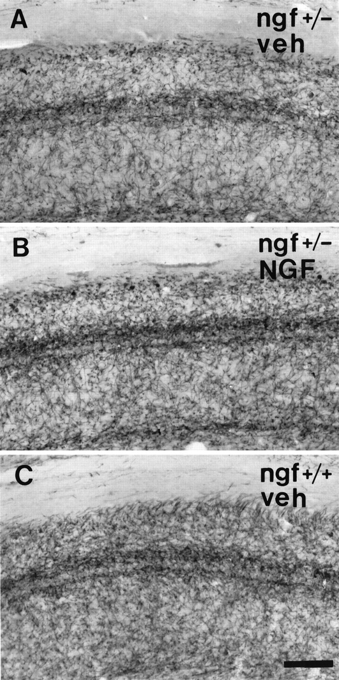
Photomicrograph of the hippocampus of (A) a ngf+/− mouse that had received vehicle, (B) a ngf+/− mouse that had received NGF, and (C) angf+/+ mouse that had received vehicle stained with AChE. Scale bar, 100 μm.

Photomicrograph of the hippocampus of (A) a ngf+/− mouse that had received vehicle, (B) a ngf+/− mouse that had received NGF, and (C) angf+/+ mouse that had received vehicle stained with AChE. Scale bar, 100 μm.