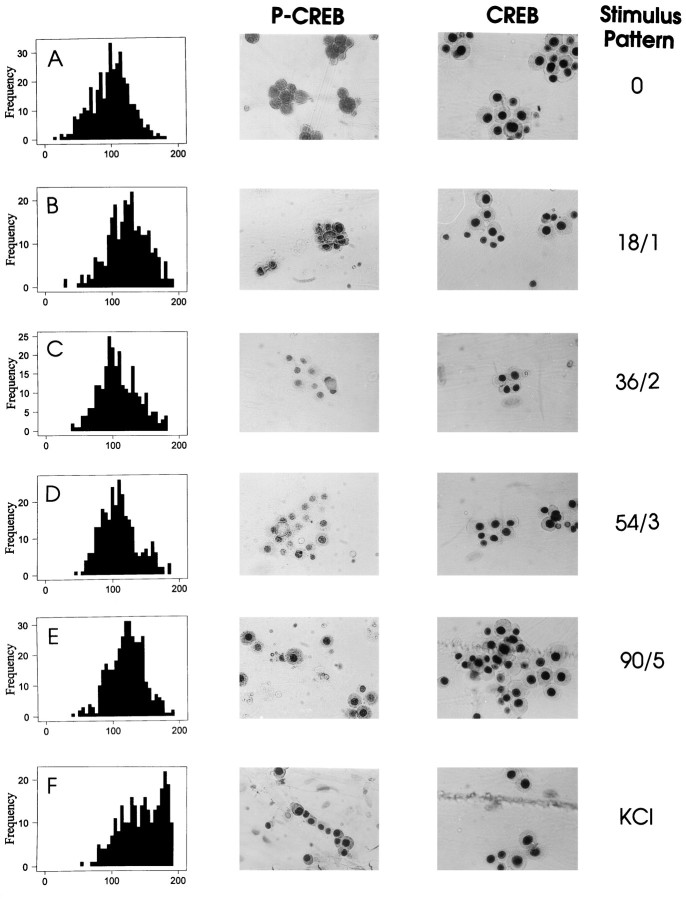
Fig. 6.
CREB phosphorylation at Ser-133 in response to electrical stimulation of different patterns. Phosphorylation was determined by nuclear staining using an antibody that recognizes CREB phosphorylated at Ser-133 (P-CREB). The intensity of immunocytochemical staining was quantified in the nucleus of stimulated cells by densitometry of digitized images on a scale of 0–255. All values were normalized to the mean intensity of nuclear staining in unstimulated cells (A). A 10 min incubation in 60 mm KCl caused a large increase in the number and intensity of nuclei staining for P-CREB (F), which is evident by the rightward shift in the histogram of nuclear staining intensities. After electrical stimulation, localization of P-CREB in the nucleus varies with different stimulus patterns (B–E). The highest levels of nuclear staining were produced by short bursts repeated frequently (1.8 sec at 10 Hz, every minute) (B) or longer duration bursts repeated infrequently (9 sec at 10 Hz, every 5 min) (E). The intermediate patterns of stimulation produced less CREB phosphorylation at Ser-133 (C, D). No change in nuclear staining was evident after any stimulus when an antibody that recognizes both the phosphorylated and dephosphorylated forms of the protein was used (A–E).

