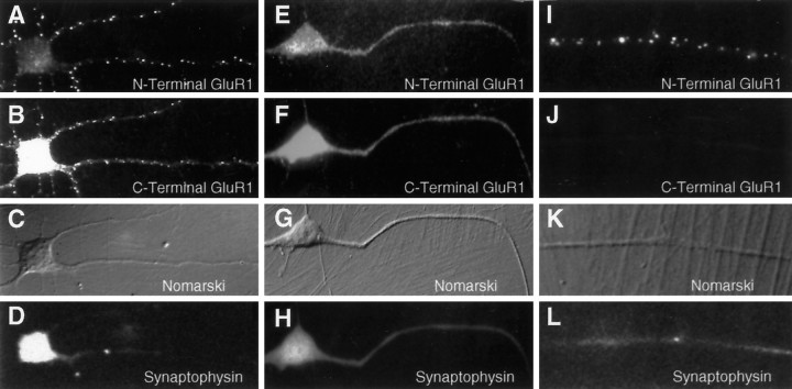
Fig. 2.
Induction of extrasynaptic receptor patches by the N-GluR1 antibody. Live, 3-d-old cultures of spinal cord neurons were incubated with whole N-GluR1 (A–D, I–L) or a Fab fragment of N-GluR1 (E–H) and then fixed and processed as described. Antibody-induced surface patches of N-GluR1 (A), which are not seen with a Fab fragment of N-GluR1 (E), have corresponding clusters of C-terminal GluR1 staining (compare B withF). In nonpermeabilized neurons (I) surface N-GluR1 staining is observed, whereas C-terminal staining is not detectable. The nonsynaptic location of this staining is confirmed by the lack of associated synaptophysin stain (D, H, L), and the absence of cell–cell contact at these sites (C, G, K).

