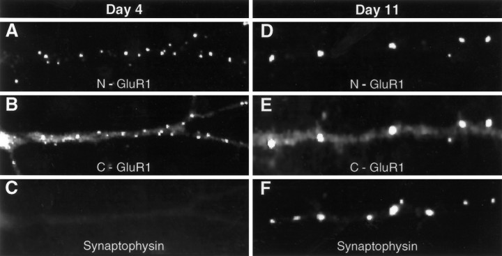
Fig. 3.
The distribution of surface GluR1 changes over time in culture. Antibody-induced, nonsynaptic patches of GluR1 are abundant on the dendrites of neurons after 4 d in culture (A–C). On day 11, however (D–F), all live cell staining with N-GluR1 is confined to synapses, defined by the presence of presynaptic synaptophysin stain (F). Note that N-GluR1-induced antibody patching appears to cluster all the surface immunostaining at both day 4 (A) and day 11 (D) but leaves a significant portion of the total GluR1 signal, seen with the C-terminal antibody, unclustered (B, E).

