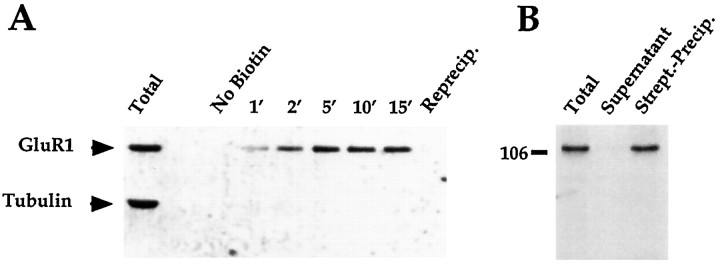
Fig. 7.
Time course and efficiency of biotinylation of GluR1 in cultured spinal cord neurons. Cultures of spinal cord neurons were biotinylated at 4°C with 1 mg/ml NHS-SS-biotin. InA, cultures were exposed to the biotinylating reagent for different times, and the detergent soluble fraction of each culture was added to streptavidin-conjugated beads and incubated for 2 hr. The supernatant recovered from the precipitation of the 15 min-treated neurons was reprecipitated with streptavidin beads, and the additional streptavidin-precipitated material was loaded as well. All samples were loaded such that each lane represents 1% of the total material per plate. The gel was transferred to immobilon and probed with both C-GluR1 and anti-tubulin antibodies. In B, one plate of neurons was scraped into the biotinylating reagent, sonicated, spun at 14,000 × g for 15 min, and resuspended in precipitation buffer including 0.2% SDS and 1% Triton X-100. A fraction of this was saved and loaded as total extract. The remainder was incubated for 2 hr with streptavidin-conjugated beads. The supernatant, streptavidin-precipitated, and total extracts were loaded such that each lane represents 1% of the material from the plate. The gel was transferred and probed with the C-GluR1 antibody.

