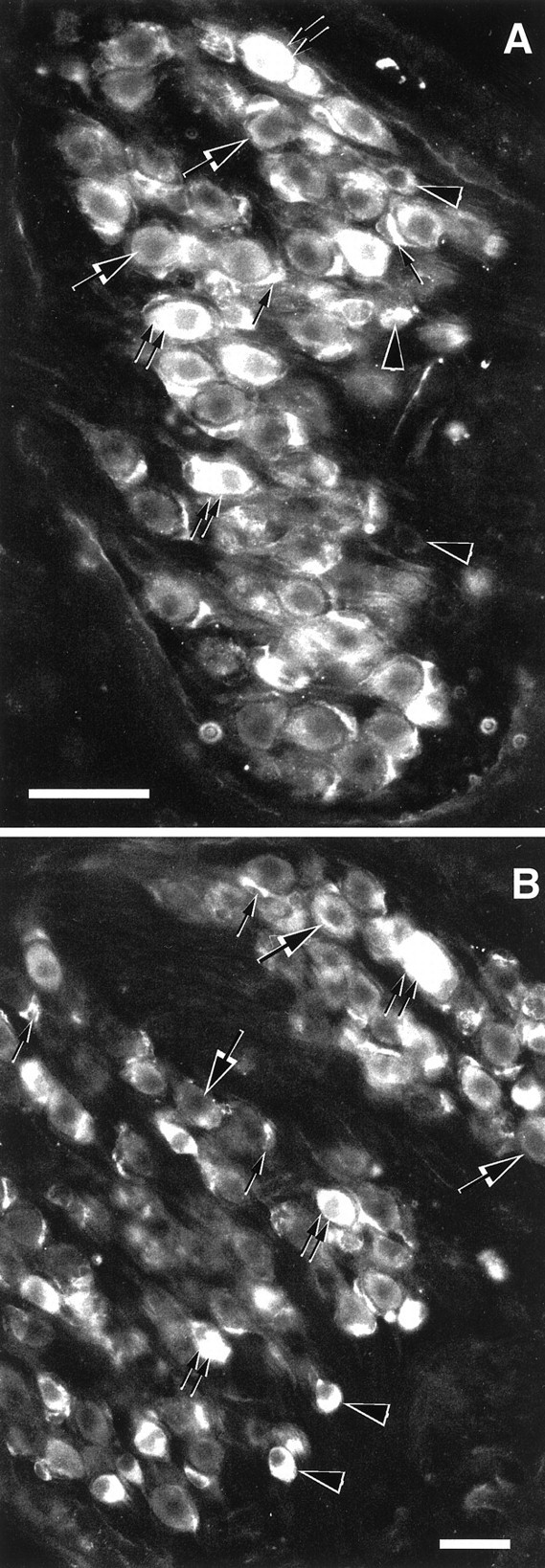
Fig. 6.

Sections through the basal turn (A) and through the apical turn (B) of the rat spiral ganglion incubated at 1.5 μg/ml with anti-δ1 and -δ1/2 antibodies, respectively. All type I (large and double arrows) and type II (arrowheads) spiral ganglion neurons show staining to both δ1 and δ1/2 antibodies. The intensity of the immunostaining is moderate (large arrows) to intense (double arrows). Satellite glial cells show intense immunostaining to δ1 and δ1/2 antibodies (small single arrows). Scale bars: A, B, 50 μm.
