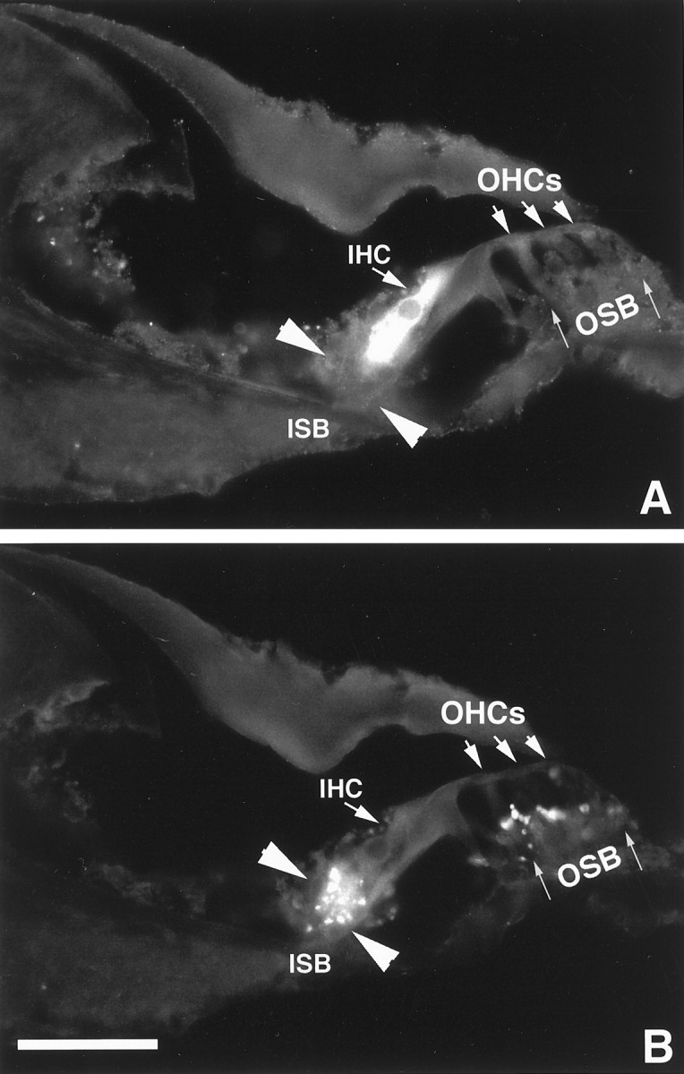
Fig. 7.

Colocalization of the immunoreactivity to δ1 (A) and synaptophysin (B) antigens in the organ of Corti of the guinea pig. A, Field illuminated for DTAF fluorescence. B, Field illuminated for rhodamine. The IHC is immunoreactive to the δ1 antibody only, and no immunostaining is seen in theOHC region (A). TheISB, delineated by two arrowheads, and the OSB, indicated by thin arrows, are stained with antibody to synaptophysin antibody (B) but not with δ1 antibody (A). Scale bar, 50 μm.
