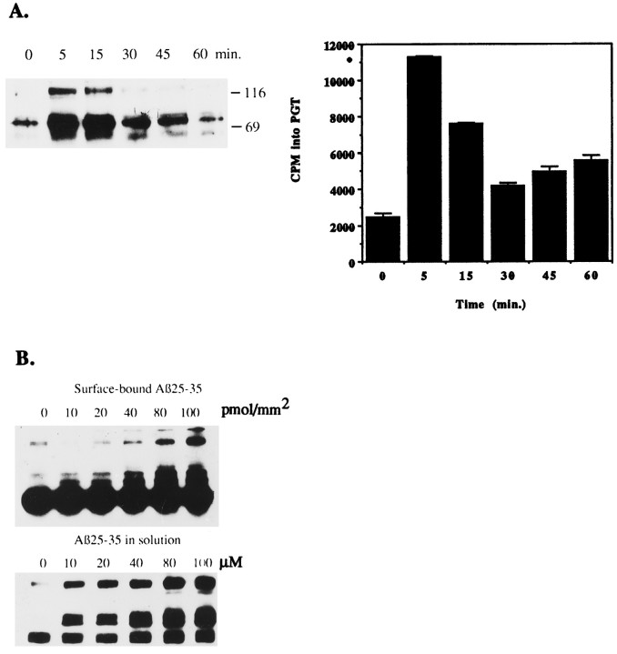
Fig. 2.
Time course and dose–response of Aβ25–35-stimulated tyrosine phosphorylation in THP1 monocytes.A, THP1 monocytes were stimulated for the indicated times on 60 pmol/mm2 Aβ25–35 bound to tissue culture dishes. Tyrosine-phosphorylated proteins were immunoprecipitated with anti-phosphotyrosine mAb (PY20), followed by elution with 40 mmp-nitrophenylphosphate. Proteins (0.6 μg) were resolved by SDS-PAGE, transferred to polyvinylidene fluoride (PVDF), and subjected to Western blot with anti-phosphotyrosine mAb (PY20). In parallel assays, proteins (0.1 μg) were analyzed in a kinase assay by phosphorylation of the tyrosine kinase substrate polyGluTyr (PGT). B, THP1 monocytes were stimulated with the indicated quantities of Aβ25–35, surface-bound or in solution, for 5 min. Tyrosine-phosphorylated proteins were immunoprecipitated with anti-phosphotyrosine mAb (PY20), resolved by SDS-PAGE, transferred to PVDF, and subjected to Western blot with anti-phosphotyrosine mAb (4G10). The broad band at 55 kDa is IgG heavy chain.

