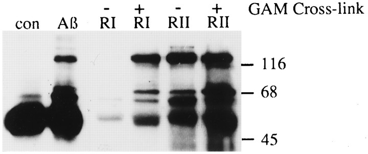
Fig. 3.
Comparison of tyrosine phosphorylation in THP1 monocytes stimulated with Aβ, FcγRI (RI), or FcγRII (RII). THP1 monocytes were stimulated for 5 min with 60 pmol/mm2Aβ25–35 bound to a tissue culture dish, mAb 32.2 (anti-FcγRI), mAb 32.2 (anti-FcγRI) cross-linked with goat anti-mouse F(ab)2, mAb IV.3 (anti-FcγRII), or mAb IV.3 (anti-FcγRII) cross-linked with goat anti-mouse F(ab)2. Tyrosine phosphoproteins were analyzed by immunoprecipitation, followed by Western blot with anti-phosphotyrosine mAb (4G10).

