
Fig. 4.
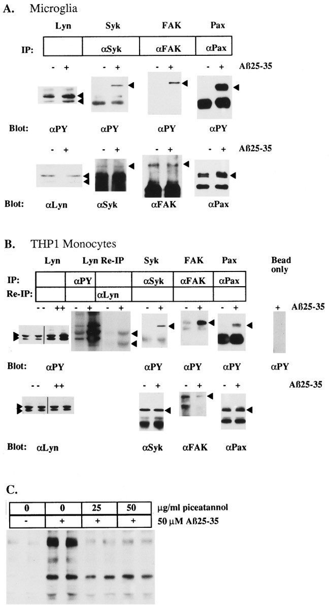
Identification of Aβ25–35-stimulated tyrosine-phosphorylated proteins in microglia and THP1 monocytes and the effect of the tyrosine kinase inhibitor piceatannol.A, Aβ25–35-stimulated tyrosine phosphorylation ofLyn, Syk, FAK, and paxillin (Pax) in primary cultures of rat microglia. B, THP1 monocytes were evaluated by immune precipitation and Western blot analysis with the indicated antibodies. Lyn identification and enzymatic activation in THP1 monocytes also was evaluated by immunoprecipitation with an anti-phosphotyrosine antibody, followed by incubation of the immune complex with [32P]ATP. The radiolabeled Lyn was released from the immune complex and then reprecipitated with an anti-Lyn antibody and visualized by autoradiography. Arrowheadsdenote migration of the respective proteins. C, THP1 monocytes were pretreated for 1 hr with the indicated amounts of piceatannol. Then cells were exposed to 50 μm Aβ25–35 for 5 min. Cellular lysates were resolved by SDS-PAGE, transferred to PVDF, and subjected to Western blot with anti-phosphotyrosine mAb (4G10).