Fig. 5.
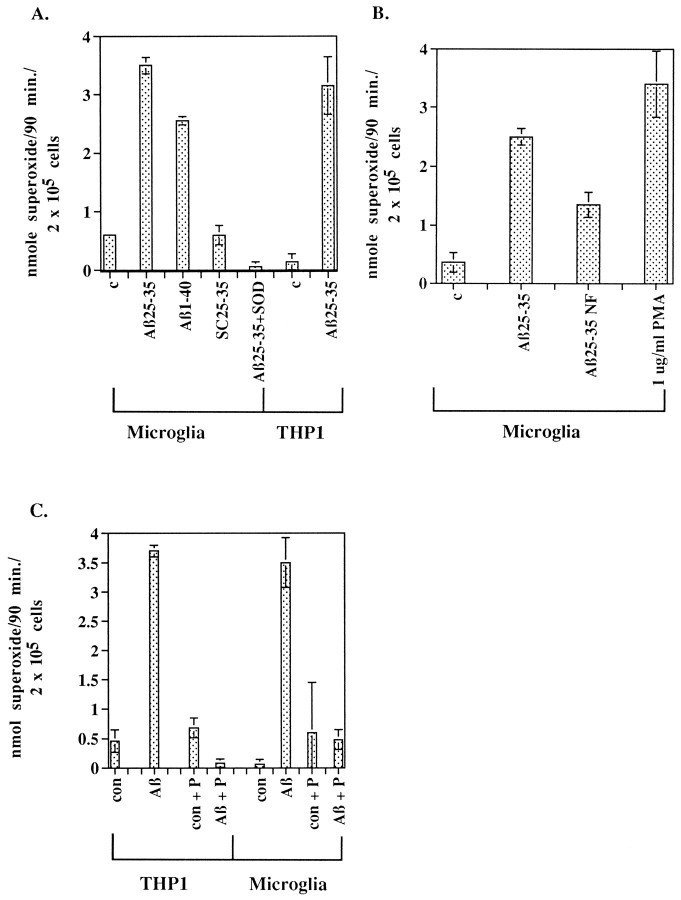
Fibrillar Aβ peptide stimulates respiratory burst in microglia and THP1 monocytes. A, Rat microglia were incubated in the absence (c) or presence of 60 μm fibrillar Aβ1–40, Aβ25–35, or scrambled Aβ25–35 (SC25–35). The peptides were added alone or in the presence of 40 μg of superoxide dismutase (SOD), and O2− production was measured. THP1 monocytes were incubated in absence (c) or presence of 60 μmAβ25–35. B, Fibrillar, but not nonfibrillar, Aβ25–35 stimulated release of O2−from rat microglia. Microglia were incubated with 40 μmfibrillar Aβ25–35 or 40 μm nonfibrillar Aβ25–35 (Aβ25–35NF), 1 μg/ml phorbol myristate acetate (PMA), or vehicle only (c) in duplicate wells for 90 min. C, Aβ25–35-stimulated release of O2− from THP1 monocytes and microglia is blocked by piceatannol. Microglia and THP1 monocytes were pretreated for 1 hr with ± 25 μg/ml piceatannol (P). Cells were unstimulated (con) or stimulated for 90 min with 60 μm Aβ25–35 ± 25 μg/ml piceatannol. Supernatants were collected, and production of superoxide was determined spectrophotometrically by measurement of the reduction of ferricytochrome C at 550 nm and converted to moles of O2−, using an extinction coefficient of 21 × 103m−1 cm−1. Production of O2− is expressed as nanomole O2− per 90 min per 2 × 10⁵cells.

