
Fig. 5.
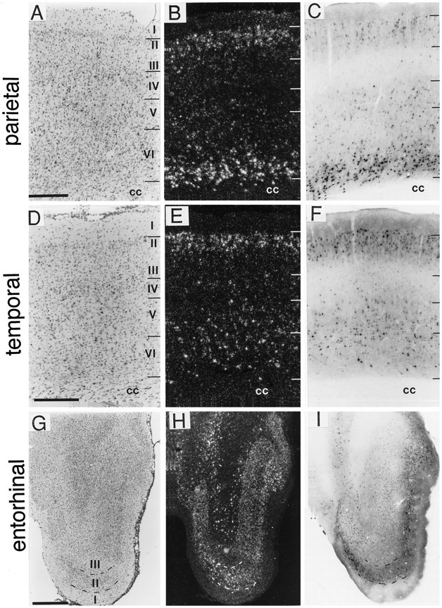
Comparison of the distributions of BDNF-ir and mRNA in cortical fields. Sections through parietal (A–C), temporal (D–F), and entorhinal (G–I) cortices were processed forin situ hybridization to localize BDNF mRNA (B, E, H; dark field) or immunohistochemistry to localize BDNF-ir (C, F, I; bright field). Sections through similar planes were Nissl-stained (A, D, G; bright field). In parietal cortex (A–C), the BDNF cRNA heavily labeled neurons in layer VI, moderately in layersII/III, and lightly in layer V; BDNF-ir was very heavy in deep layer VI, lighter in layersII, III, V, and superficial layer VI, and almost undetectable in layer IV. In temporal cortex (D–F), BDNF mRNA was moderately labeled in layers II/III and V/VI, with lighter labeling in layer IV. BDNF-ir in temporal cortex was heaviest in layers II/III, moderate in layersV/VI, and light in layer IV. In entorhinal cortex (G–I), the patterns of BDNF cRNA hybridization and immunoreactivity were similar. Scale bars (shown in A): A–C, 500 μm; (shown inD): D–F, 500 μm; (shown inG): G–I, 1 mm. cc, Corpus callosum.