Fig. 6.
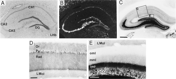
Distribution of BDNF-ir and mRNA in hippocampus. Low-magnification (A–C) and high-magnification (D, E) photomicrographs of sections through hippocampus that were either Nissl-stained (A, bright field) or processed for in situ hybridization (B, dark field) or immunohistochemistry (C–E, bright field). In hippocampus, the BDNF cRNA densely labeled pyramidal cells in CA3, CA2, and the hilus, and less densely the CA1 pyramidal cells and the dentate gyrus granule cells (GrDG) (B). BDNF immunostaining densely labeled processes within the mossy fiber system (C). At higher magnification, one could see that in region CA1 BDNF immunostaining was diffuse in strata oriens (Or) and radiatum (Rad) and weaker in stratum lacunosum moleculare (LMol) (D). Scattered neurons in the CA1 pyramidal cell layer (Py) were immunolabeled (D). In the dentate gyrus (DG) molecular layer, a trilaminar pattern of BDNF-ir was seen, with moderately dense labeling in the inner molecular layer (iml), light labeling in the middle molecular layer (mml), and no detectable staining in the outer molecular layer (oml). Scale bars (shown in C):A–C, 500 μm; (shown in D): D, E, 100 μm and 75 μm, respectively. LHb, Lateral habenular nucleus.

