
Fig. 8.
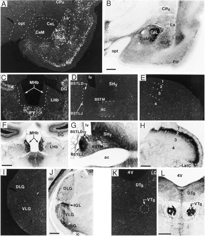
Dense fiber/terminal BDNF-ir was present in many regions lacking BDNF mRNA expression. A–L, Dark-field and bright-field photomicrographs of sections processed for in situ hybridization (A, C–E, I, K) and immunohistochemistry (B, F–H, J, L), respectively, through the amygdala (A, B), habenula (C, F), bed nucleus of stria terminalis (D, G), spinal cord (E, H), lateral geniculate nucleus (I, J), and dorsal/ventral tegmental nucleus (K, L). As seen in Aand B, in the basolateral (BLA) and lateral amygdala (La), many cells were heavily labeled for both mRNA and BDNF-ir, whereas in the central amygdala many immunolabeled fibers/terminals were present but cRNA hybridization was not. Similarly, in the medial habenular nucleus (MHb) (C, F) subregions of the bed nucleus of stria terminalis (D, G), the dorsal (DLG) and ventral regions (VLG) of the lateral geniculate nucleus (I, J), the intergeniculate leaf (IGL) (I, J), and ventral tegmental nucleus (VTg) (K, L), heavy fiber/terminal BDNF-ir was found without detectable mRNA. In spinal cord (cervical level shown), heavily immunostained fibers were present primarily in lamina 2 (H), which lacked BDNF mRNA (E). Scale bars (shown in B): A, B, 500 μm; (shown in F): C, D, F, G, 500 μm; (shown in H): E, H, 150 μm; (shown in J, L):I–L, 500 μm. ac, Anterior commissure; BSTLD, bed nucleus of stria terminalis, lateral dorsal; BSTLJ, bed nucleus of the stria terminalis, juxtacapsular; BSTM, bed nucleus of stria terminalis, medial; CeL, central amygdaloid nucleus, lateral division; CeM, central amygdaloid nucleus, medial division; CPu, caudate putamen (striatum); DEn, dorsal endopiriform nucleus;DG, dentate gyrus; DTg, dorsal tegmental nucleus; HF, hippocampal formation; ic, internal capsule; LatC, lateral cervical nucleus;LC, locus coeruleus; LHb, lateral habenular nucleus; lv, lateral ventricle;opt, optic tract; Pir, piriform cortex;PV, paraventricular thalamic nucleus;SHy, septohypothalamic nucleus; SubG, subgeniculate nucleus; 4V, 4th ventricle.