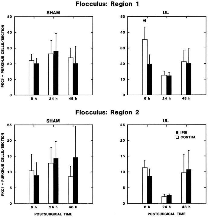
Fig. 11.
Transient regional changes in flocculus Purkinje cell expression of PKCδ. The number of PKCδ-immunopositive Purkinje cells per section from every sixth section through the intermediate aspect of flocculus is plotted as a function of postoperative time. Increased expression was observed contralaterally 6 hr after UL (Newman–Keuls test; p < 0.01) in region 1, which included the entire ventral surface and intermediate third of the dorsal surface of the nodulus. No significant effects were observed in region 2, the lateral and medial thirds of the dorsal surface of the flocculus.

