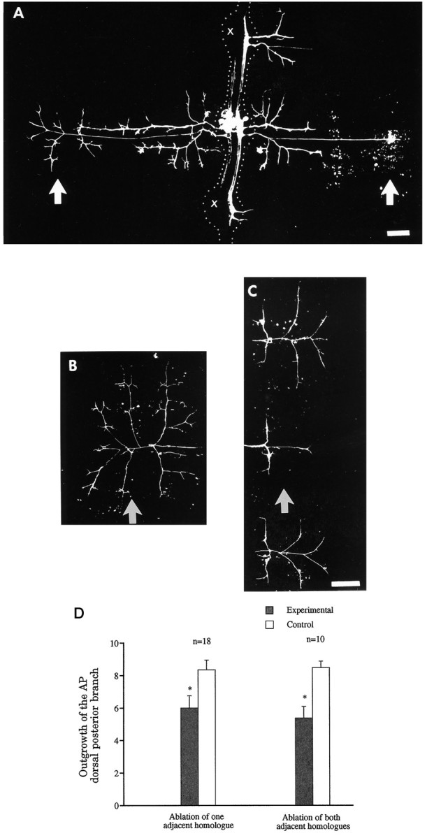
Fig. 7.

Greater outgrowth of its longitudinal projections reduces the arborization of the lateral projections of the AP cell.A, In this preparation, four AP cells were ablated at E9–E10 on the same side in the two ganglia adjacent to the ganglion of interest (crosses) as well as the next two ganglia (not shown). Two days later, the experimental AP cell on the same side as the ablated AP cells had extended its longitudinal projections ectopically to the periphery, through the adjacent ganglia (on theright of the picture). At the same time, the lateral projections of this AP cell grew less in its own segment than those of its contralateral homolog, which serves as the control. The effect is more obvious in the dorsal germinal plate (arrows).Dotted lines were added to show boundaries of ganglia and interganglionic connectives. B,C, Another example, 4 d after ablation of the four adjacent ipsilateral AP cells, of the difference in outgrowth of control (B) and experimental (C) AP cells in the dorsal germinal plate (corresponding to the region in A indicated byarrows). The longitudinal projections of the experimental AP cell had grown extensively in the dorsal region of the adjacent segments (panel C), but the lateral projection had significantly reduced outgrowth in the dorsum of its own segment (arrow), compared with the contralateral homolog shown in B (arrow). D, Quantitative measurements of the arbors of control and experimental AP cells in the dorsal germinal plate of their own segments. The data, taken 2 d after ablating either one AP cell located ipsilaterally in either an anterior or a posterior adjacent ganglion (left) or both adjacent ipsilateral AP cells (right), show 35 and 50% reductions in outgrowth, respectively (asterisks). Anterior is at thetop in panels A–C. Scale bars, 100 μm; scale bar in C also applies to B.
