
Fig. 5.
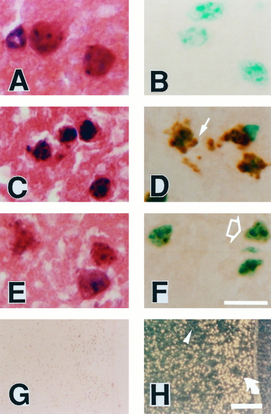
Representative findings of TUNEL staining in Wt mice at 24 hr after ischemia. High magnification of neuronal cells in the caudate putamen with H & E staining (A,C, E) and TUNEL staining with methyl green counter staining (B, D,F). A, B, The nuclear and cellular morphology of the neurons in the nonischemic caudate putamen is typical of normal neurons with smooth nuclear membrane and uniform chromatin formation (A). TUNEL staining does not label normal neuronal cells (B).C, D, Representative morphology of the cells in the marginal zone of the infarction, which shows apoptotic features such as cell shrinkage and chromatin condensation in the nuclei (C). Apoptotic neuronal cells in the marginal zone are recognized with TUNEL staining with markedly labeled small particles, called “apoptotic bodies,” around the nuclei (D, arrow). E,F, Representative morphology of the cells in the infarction, which shows necrotic features such as cellular swelling (pale color on eosinophilic staining) and irregularly shaped nuclei (E). These neuronal cells are slightly labeled with TUNEL staining in the nuclei (F, open arrow).G, H, Lower magnification of TUNEL staining by bright-field (G) and dark-field (H) phase-contrast microscopy. Two patterns of the staining quality are emphasized in the dark-field phase-contrast photomicrographs: apoptotic neurons with dense TUNEL labeling asbright yellow spots (H, rotated arrow) and necrotic neurons with slight TUNEL labeling as faint, small, yellow spots (H,arrowhead). Scale bars:A–F, 20 μm; G,H, 200 μm.