
Fig. 5.
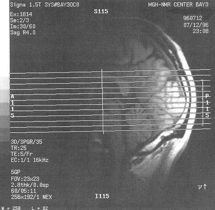
Midsagittal anatomical image from subject S1 showing the typical placing of the 12 slices used in this study. Slices were selected so as to include the entire ventral surface of the occipital and temporal lobes.
Official websites use .gov
A
.gov website belongs to an official
government organization in the United States.
Secure .gov websites use HTTPS
A lock (
) or https:// means you've safely
connected to the .gov website. Share sensitive
information only on official, secure websites.
Midsagittal anatomical image from subject S1 showing the typical placing of the 12 slices used in this study. Slices were selected so as to include the entire ventral surface of the occipital and temporal lobes.