
Fig. 2.
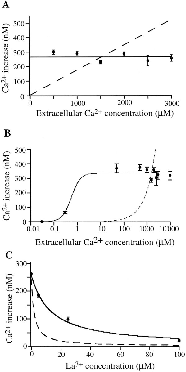
Dependence of somatic Ca2+ responses on extracellular Ca2+ concentration. A, The action potential-induced intracellular Ca2+ rise is independent of external Ca2+ concentration from 500 to 3000 μm (data points and solid line). Thedashed line illustrates the expected results of this experiment under the assumption that the intracellular Ca2+rise was the direct result of Ca2+ influx from voltage-gated Ca2+ channels. B, Only when external [Ca2+] is lowered below 50 μm does the action potential-induced Ca2+ rise become diminished.C, Dependence of somatic Ca2+ responses on extracellular lanthanum concentration. La3+ suppressed the action potential-induced intracellular Ca2+ rise but with a much higher IC50 (13 μm) than expected. An extracellular [Ca2+] of 1.5 mm was present throughout. The dashed line again represents the expectation based on the assumption of the Ca2+ rise coming directly from influx via voltage-gated Ca2+ channels, modeled on data from the effect of La3+ on individual voltage-gated Ca2+ channels, with an IC50 of 1.7 μm. The solid line is the best fit to the data with the form: A =aIC50/([La3+] + IC50), where IC50 = 13 μm, anda = 262 nm.