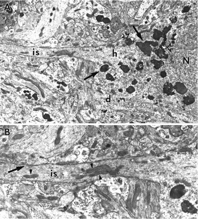
Fig. 4.

A, Electron micrograph of the basal portion of a CA1 pyramidal cell soma from a ZPAD-treated slice. Although numerous lysosomes (arrows) encircle the nucleus (N) and are found within the proximal span of a basal dendrite (d), none are observed in the axon hillock (h) and initial segment (is) within this section. Vacuole-containing lysosomes (open arrows) are also evident within the perikaryal cytoplasm. Magnification, 8300×. B, Enlargement of the axon initial segment (is). Note the dense axolemmal undercoating, microtubule bundling, cisternal organelle (arrow), mitochondria (arrowheads), and paucity of lysosomes. Magnification, 15,000×.
