
Fig. 7.
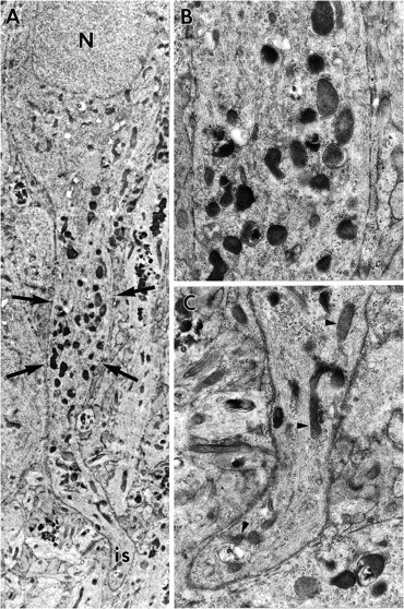
A, Montage of electron micrographs of a CA1 neuron 7 d after the conclusion of ZPAD exposure. Although the nucleus (N), soma, and axon initial segment (is) appear normal, the location of the axon initial segment is distally displaced by a fusiform expansion (arrows) of the axon hillock region (meganeurite). The cytosol of the meganeurite, unlike that of the axon and soma, contains many lysosomes. Magnification, 6200×. B, Enlargement of the indicated portion of the fusiform expansion. Magnification, 18,500×. C, Enlargement of the region containing the axon initial segment. Note that this region contains several mitochondria (arrowheads) and a few lysosomes. Magnification, 18,500×.