
Fig. 2.
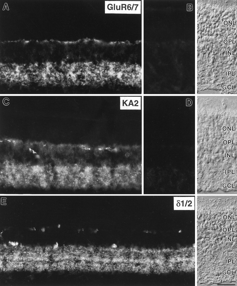
Micrographs of vertical cryostat sections of rat retina immunostained with the antisera against GluR6/7, KA2, and δ1/2. A, Diffuse and punctate labeling for GluR6/7 was present in both synaptic layers, the OPL and theIPL. B, Preadsorption of the anti-GluR6/7 antiserum with the immunogen resulted in a complete loss of specific immunoreactivity.C, Diffuse and punctate labeling for KA2 was also present in both plexiform layers of the rat retina. In addition, somata in the inner nuclear layer showed weak KA2 immunostaining.D, No specific immunolabel was detected after preadsorption of the anti-KA2 antiserum with the immunogen.E, Punctate labeling for δ1/2 was present in several distinct strata in the IPL. Stained profiles in theOPL are unspecifically labeled blood vessels. The retinal layers are shown with Nomarski optics. ONL, Outer nuclear layer; OPL, outer plexiform layer;INL, inner nuclear layer; IPL, inner plexiform layer; GCL, ganglion cell layer. Scale bar, 25 μm.