Fig. 6.

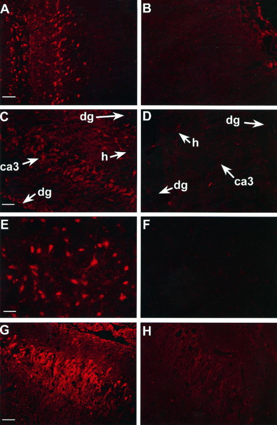
Immunofluorescent images of bcl-2 protein in brain sections 24 hr after TBI or sham-operation (bcl-2 labeling =red). bcl-2 immunoreactivity is increased in neurons in cortex, CA3 hippocampus, dentate gyrus, and hilus after TBI versus sham-operation. A, Ipsilateral cortex after TBI.B, Ipsilateral cortex after sham-operation.C, Ipsilateral hippocampus after TBI. D, Ipsilateral hippocampus after sham-operation. E,F, Consecutive brain sections incubated with (E) and without (F) primary antibody. G, H, Consecutive brain sections incubated with primary antibody (G) or primary antibody preabsorbed with rat bcl-2 peptide (H). ca3, CA3 hippocampus;dg, dentate gyrus; h, hilus. Scale bars:A–D, 50 μm; E, G, 25 μm; G, H, 100 μm.
