
Fig. 9.
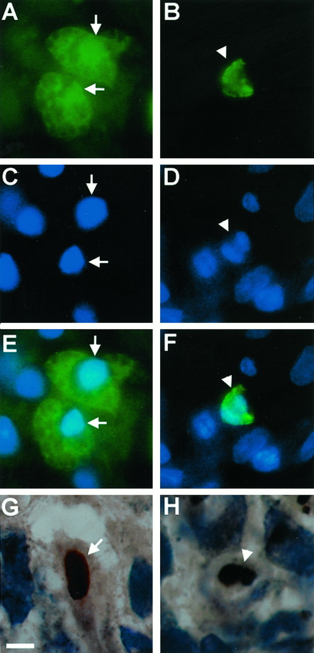
Immunofluorescent images and photomicrographs showing nuclear morphologies characteristic of both necrosis (A, C, E, G) and apoptosis (B, D, F, H) after TBI (TUNEL-FITC = green; TUNEL-diaminobenzidine = brown; bis-benzimide = blue). Note TUNEL-positive cells demonstrating a condensed, pyknotic, and clumped nuclear pattern suggestive of apoptosis (arrowheads) and TUNEL-positive cells with a diffuse pattern of TUNEL staining without shrinkage of the nuclei and with occasional extravasation of TUNEL labeling into the cytoplasm, suggestive of necrosis (arrows). A, C, E, G, Ipsilateral CA3 hippocampus at 24 hr. B, D, F, Ipsilateral cortex at 24 hr. H, Ipsilateral dentate gyrus at 24 hr. Scale bar, 10 μm.