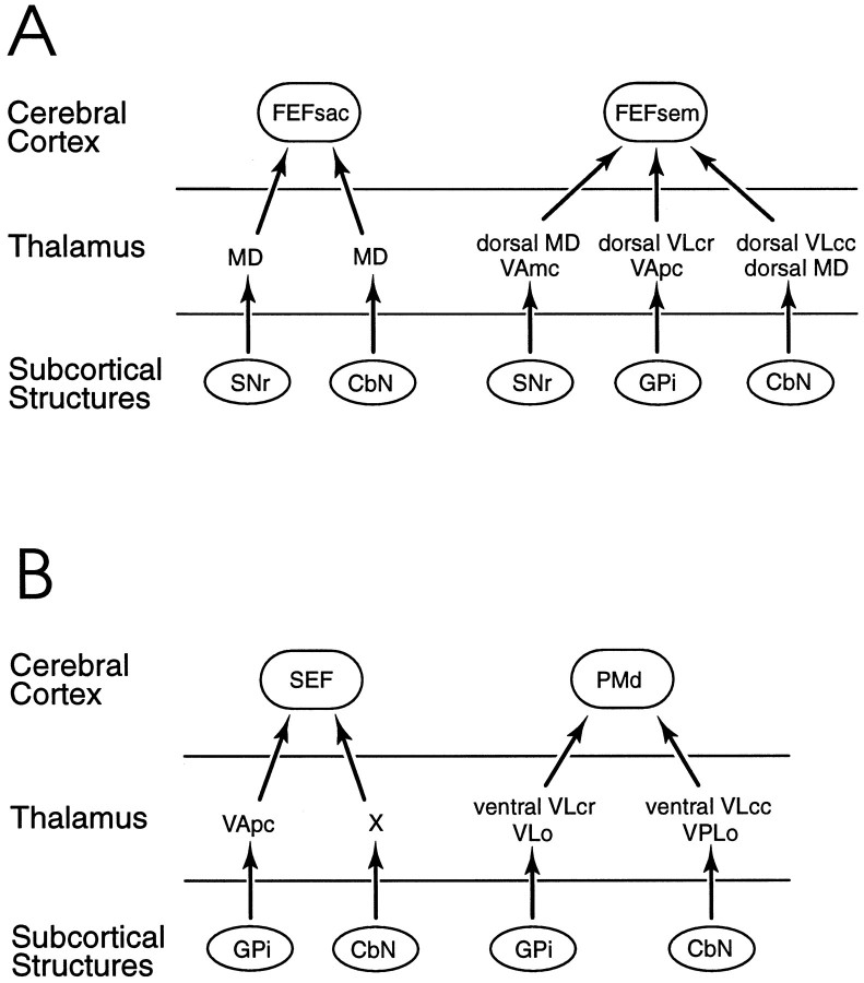
Fig. 10.
Summary diagram of GPi- andSNr-thalamocortical and cerebellothalamocortical connection patterns. A, Putative circuits from basal ganglia and cerebellum through thalamic nuclei to theFEFsac and FEFsem. B, Putative circuits from basal ganglia and cerebellum through thalamic nuclei to the SEF and PMd. Each of the functional areas in the cerebral cortex receives a major neural input from both a basal ganglia-receiving and a cerebellar-receiving cell group in the thalamus. It is proposed that some neurons from the basal ganglia and cerebellar nuclei synapse on thalamic neurons that, in turn, project to the cortical eye fields. However, this specific connectivity has thus far been confirmed with transneuronal transport experiments only in the case of the FEFsac (Lynch et al., 1994). The terms “dorsal” and “ventral” are used with theVLcr and VLcc nuclei to emphasize the fact that even though both the FEFsem and thePMd receive input from these two nuclei, the respective pathways originate in separate subregions of these nuclei. Similarly, the term “dorsal MD” is used to emphasize that theMD projection to the FEFsem originates in the dorsal-most portion of paralaminar MD, whereas theMD projection to the FEFsac originates relatively more ventrally in paralaminar MD.CbN, Cerebellar nuclei; FEF, frontal eye field; FEFsac, saccadic subregion of theFEF; FEFsem, smooth eye movement subregion of the FEF; GPi, internal globus pallidus; MD, medialis dorsalis;PMd, dorsal premotor cortex; SEF, supplementary eye field; SNr, substantia nigra, pars reticulata; VAmc, ventralis anterior, pars magnocellularis; VApc, ventralis anterior, pars parvocellularis; VLcc, caudal portion of ventralis lateralis, pars caudalis; VLcr, rostral portion of ventralis lateralis, pars caudalis; VLo, ventralis lateralis, pars oralis; VPLo, ventralis posterior lateralis, pars oralis; X, area X in the ventral lateral complex.

