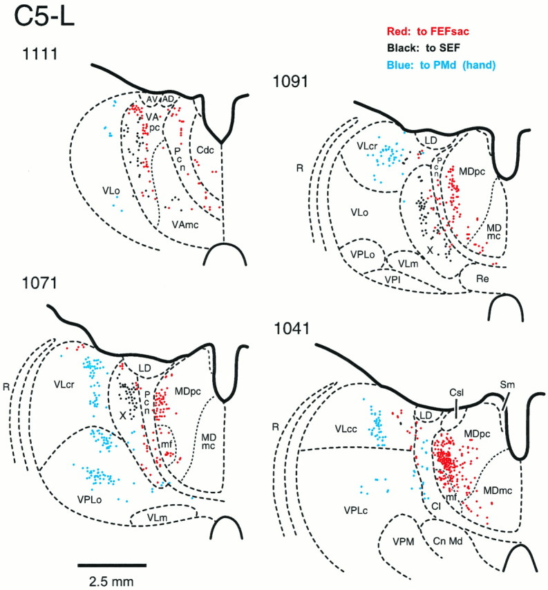
Fig. 7.

The origin of thalamic inputs to theFEFsac, SEF, and PMd in the left hemisphere of monkey C5 on coronal sections (C5-L) (see Table 1 for fluorescent tracers used in these injections). Section 1111 is at the most rostral level, and section 1041 is at the most caudal level. A total of 31 sections at 250 μm intervals were plotted. Red dots indicate the neurons labeled from an injection site in theFEFsac; black dots indicate the neurons labeled from an injection site in the SEF; andlight blue dots indicate the neurons labeled from an injection site of the PMd (hand). AD, Anterior dorsalis; AV, anterior ventralis;Cdc, centralis densocellularis; Cl, central lateral nucleus; Cn Md, centrum medianum;Csl, centralis superior lateralis; LD, lateralis dorsalis; MDmc, medialis dorsalis, pars magnocellularis; mf, medialis dorsalis, pars multiformis; MDpc, medialis dorsalis, pars parvocellularis; Pcn, paracentral nucleus;R, reticular nucleus; Re, nucleus of reuniens; Sm, stria medullaris thalami;VAmc, ventralis anterior, pars magnocellularis;VApc, ventralis anterior, pars parvocellularis;VLc, ventralis lateralis, pars caudalis;VLcc, caudal portion of VLc;VLcr, rostral portion of VLc;VLm, ventralis lateralis, pars medialis;VLo, ventralis lateralis, pars oralis;VPI, ventralis posterior inferior; VPLc, ventralis posterior lateralis, pars caudalis; VPLo, ventralis posterior lateralis, pars oralis; VPM, ventralis posterior medialis; X, area Xin the ventral lateral complex.
