
Fig. 1.
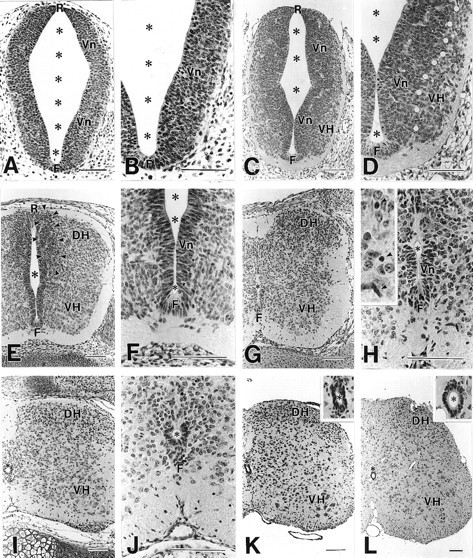
Histology of the developing spinal cord at E9 (A, B), E11 (C, D), E13 (E, F), E15 (G, H), E18 (I, J), P7 (K), and P21 (L). Hematoxylin-stained paraffin sections.White circles in D indicate the border between the ventricular zone (Vn) and ventral horn (VH). Arrowheads inE indicate a transient cell-dense layer formed between the ventricular zone and dorsal horn (DH).Arrows in H indicate putative glia cells with small dark nuclei. F, Floor plate;R, roof plate; *, central canal. Scale bars, 100 μm.