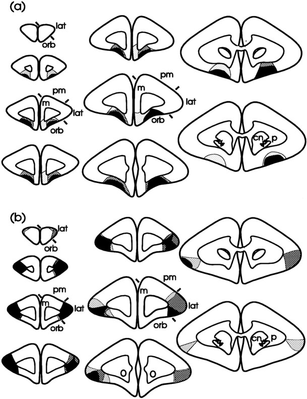
Fig. 4.
Schematic diagrams of a series of coronal sections through the frontal lobe illustrating the site of the lesion of the orbital (a) and lateral (b) prefrontal cortices. The three different types of shading represent the area of tissue that was damaged in all three marmosets (black shading), in two of the three marmosets (dark stippling), and in one marmoset only (pale stippling) after an orbital (a) or a lateral lesion (b). Orbital prefrontal cortex corresponds to areas 10–13 (marked orb on the sections), and lateral prefrontal cortex corresponds to area 9 (markedlat on the sections), as defined by Brodmann (1909) in his description of the frontal cortex in the marmoset.m, Medial prefrontal cortex; pm, premotor cortex; cn, caudate nucleus; p, putamen).

