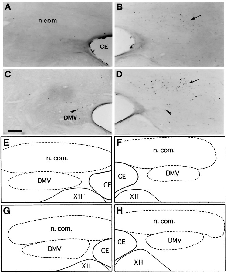
Fig. 5.
Bright-field photomicrographs of transverse sections illustrating FLI in the left (A, C) and right (B, D) commissural subdivision (n com) of the nTS. Control (A), coughing (B), codeine-control (C), and stimulated–treated (D) cats. FLI was induced in n.com of the two cats subjected to SLN stimulation whether or not they were treated with codeine (arrows in B andD). Codeine induced a few Fos-positive neurons in the dorsal motor nucleus of the vagus nerve (DMV) (arrowheads in C and D).E, F, G, and H are schematic drawings of dorsal medullary nuclei shown in A, B, C, andD, respectively. Outlined structuresindicate selected areas in which labeled cells were counted. Scale bar, 220 μm. CE, Central canal; XII, hypoglossal nucleus.

