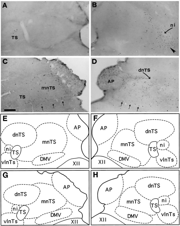
Fig. 6.
Bright-field photomicrographs of transverse sections illustrating FLI in the dorsal vagal complex (nTS, AP, and DMV) of medulla. Control (A), coughing (B), codeine-control (C), and stimulated–treated (D) cats. Note dense labeling in the interstitial (ni, curved arrow) and ventrolateral (arrowhead) subdivisions of the nTS observed only inB. Codeine induced sparse Fos-positive neurons in AP, DMV (three small arrows), and mnTS of codeine-control cat (C). Similar FLI was also observed in AP and DMV of the stimulated–treated (D) cat, associated with an increase in Fos-like expression in the dnTS and mnTS. E, F, G, and H are schematic drawings of dorsal medullary nuclei shown in A, B, C, and D, respectively. Outlined structuresindicate selected areas in which labeled cells were counted. Scale bar, 300 μm. Abbreviations are defined in legend to Figure 3.

