
Fig. 1.
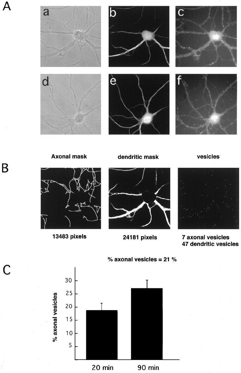
Dendritic and axonal distribution of internalized Tf. A, Hippocampal neurons were incubated for 2 hr in medium without Tf followed by incubation in 300 nm Rh-Tf for 20 min (a–c) and 90 min (d–f). a, d, Phase contrast;b, e, MAP2 labeling (dendritic marker); c, f, internalized Tf. After 20 min of internalization (c) Tf positive structures are preferentially dendritic (compare the distribution of dots inc with that of the MAP2-positive processes inb). After 90 min of internalization (f), numerous dots are present in processes (small arrows inf) negative for MAP2 (e).B, Example (based on the cell shown in A, a–c) of how the computer program identifies axons and dendrites and determines the distribution of Tf-positive (rhodamine) dots. The computer recognizes dendrites on the basis of MAP2 labeling (middle panel) and axons by phase contrast (left panel) and lack of MAP2 labeling, and then allocates and counts the Tf dots. C, With longer incubation times there is an increase in Tf-positive structures in axons.