
Fig. 3.
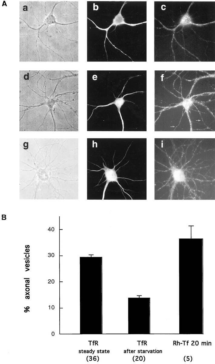
The distribution of the receptor changes after ligand addition. A, Hippocampal neurons were incubated for 2 hr in medium without Tf (a–c) or with 300 nm holo-Tf (d–f). The cells were then fixed, permeabilized, and double-labeled with an antibody against the TfR (c, f) and the dendritic marker MAP2 (b, e). Without Tf in the medium the receptor is preferentially dendritic (compare the distribution of the TfRdots in c with that of MAP2-positive dendrites in b). After ligand addition, several dots are evident in axons, as evidenced by their lack of MAP2 staining (e). g–i, Nonstarved cells (as ind–f) incubated for 20 min with Rh-Tf (i), fixed, and analyzed by fluorescence microscopy. Even after 20 min of internalization, axonal positive structures are seen. Rh-Tf dots are abundant in both axons and dendrites (compare with the pattern of MAP2 labeling of this cell inh). B, In the presence of Tf in the medium (TfR steady state) the number of TfR-positive structures in axons increases more than twofold with respect to cells starved for 2 hr (absolute values and significance are given in text). The number of cells analyzed is in parentheses.