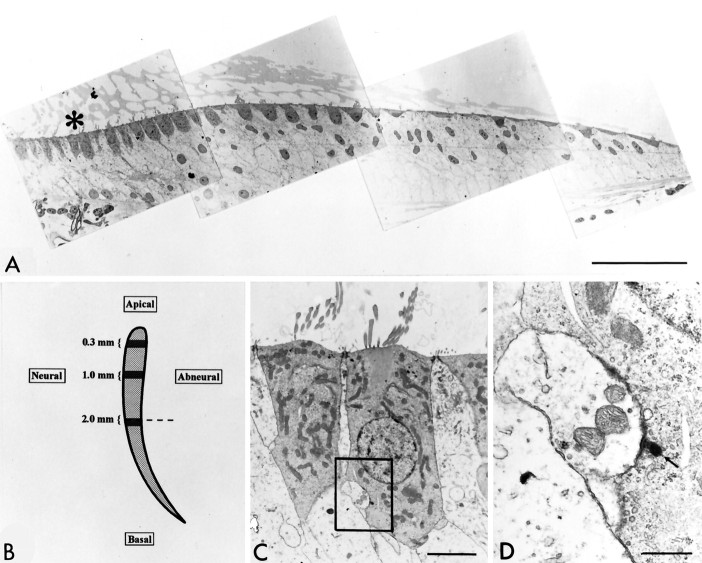
Fig. 1.
Mapping of release sites in the basilar papilla.A, Thin section electron micrographic montage from the 2 mm region of the basilar papilla. Hair cells are characteristically darker than are supporting cells. The asterisk is positioned at the apical surface of two neural (tall) hair cells that are viewed at higher magnification in C andD. Scale bar, 57 μm. B, Schematic representation of the chick basilar papilla. The three regions in which cells were studied are highlighted and labeled. Dashed line through the 2 mm zone indicates the approximate location and orientation of the montage in A. C, Neuronal hair cells from the 2 mm zone (indicated by theasterisk in A) were photographically enlarged. An afferent synapse with a PDB is identified in the box. Scale bar, 3.6 μm. D, PDB (arrow) and afferent terminal in 2 mm neural hair cell featured inC. Scale bar, 0.7 μm.

