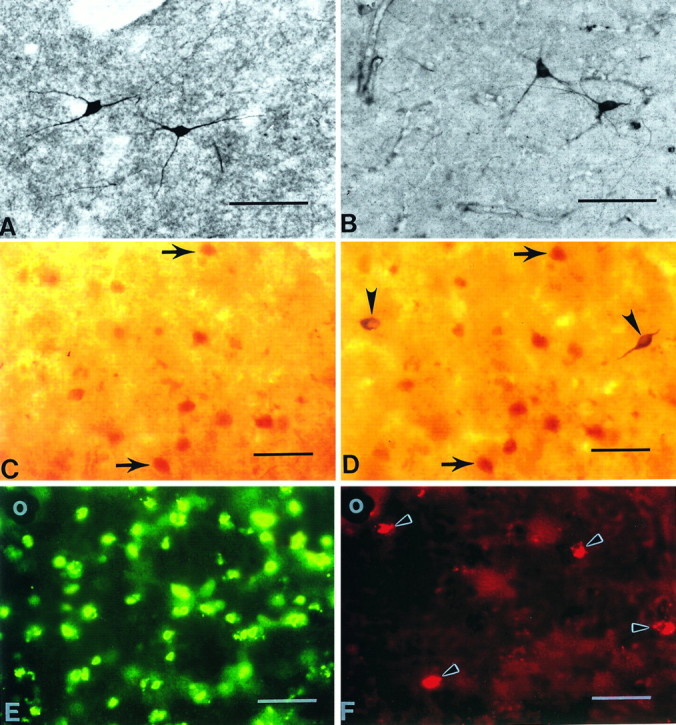
Fig. 7.

Comparison of huntingtin-positive neurons with NADPH-diaphorase (NADPH-d)-positive and nitric oxide synthase (NOS)-positive neurons in the normal caudate nucleus. Striatal NADPH-d neurons (A) and NOS neurons (B) are morphologically similar and are reported to colocalize (Hope et al., 1991). A striatal caudate section, first immunostained for huntingtin immunoreactivity (C) and subsequently treated for NADPH-d enzyme histochemistry (D), suggests that NADPH-d neurons do not contain huntingtin. Arrows inC and D delineate the same huntingtin-positive neurons within the section. There are no corresponding huntingtin-positive neurons in C where NADPH-d neurons are observed in D(arrowheads). Combined immunofluorescence for huntingtin (FITC) (E) and NOS (TRITC) (F) immunoreactivities in the same section confirm the absence of huntingtin and NADPH-d colocalization found in C andD. NOS-positive neurons in F(arrowheads) do not correspond with any huntingtin neurons in E. The blood vessel in the top left corner of E and F (white circles) acts as a fiduciary mark. Magnification bars inA–F, 100 μm.
