
Fig. 1.
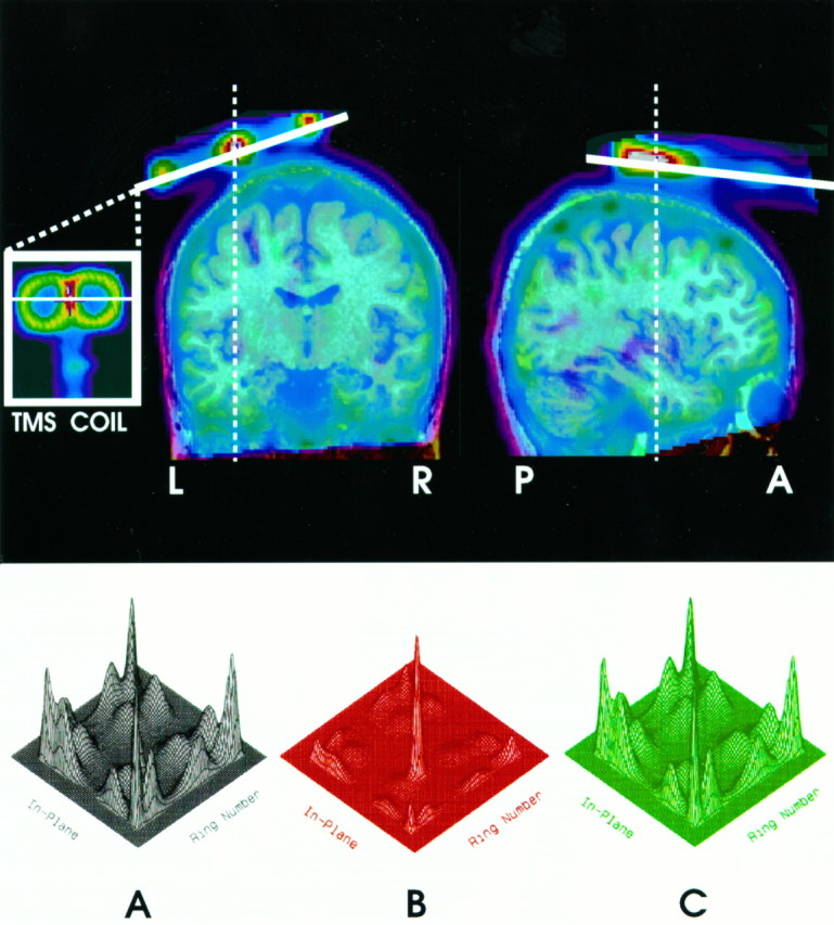
The top of the figure shows a coronal (left) and a sagittal (right) section through a transmission scan obtained in one subject, superimposed on an MR image of the same subject. The TMS coil can be seen in the inset. Note the figure-eight shape of the coil. The bottom of the figure contains three-dimensional plots of the crystal identification matrix obtained in the absence of magnetic field (A), during magnetic stimulation (B), and during the same magnetic stimulation but with metal shields placed between the coil and the photo multipliers (C). Note a serious distortion of the matrix during unshielded exposure to the pulse magnetic field (B).