Fig. 3.
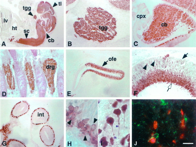
Distribution of Hu immunoreactivity in the E14 and adult mouse. A, Sagittal section (11 μm) from E14 mouse stained with affinity-purified Hu antiserum. There is intense immunoreactivity in the central and peripheral nervous systems, including the telencephalon, cerebellum, and spinal cord, but no reactivity in other tissues, including liver and heart.B, Hu immunoreactivity in E14 trigeminal ganglia.C, Hu immunoreactivity in E14 cerebellum, demonstrating intense staining in the developing cerebellum with no staining of the choroid plexus. D, Hu immunoreactivity in E14 dorsal root ganglia. E, Hu immunoreactivity in E14 olfactory epithelium. F, Hu immunoreactivity in E14 retina; Hu staining is intense in ganglion cell layer (open arrow) but absent in the ventricular surface (solid arrow), except in some scattered cells (arrowheads).G, Hu expression in the ganglion cells in the small intestine. H, High-power magnification of Hu immunoreactivity in E14 cortex. Note the cytoplasmic staining in the developing cells (arrowheads). I, High-power magnification of Hu immunoreactivity in a horizontal section (11 μm) of adult cortex, demonstrating that Hu reactivity in the differentiated neuron is both nuclear and cytoplasmic (arrows). J, Immunofluorescence double exposure of GFAP (green) and Hu(red) immunoreactivity in a horizontal section of adult cortex, demonstrating that the two are mutually exclusive.tl, Telencephalon; tgg, trigeminal ganglia; sc, spinal cord; cb, cerebellum;ht, heart; lv, liver; cpx, choroid plexus; drg, dorsal root ganglia;ofe, olfactory epithelium; int, small intestine. Scale bars: 2 mm in A; 160 μm inB–E, G; 80 μm in F; 20 μm in H–J.

