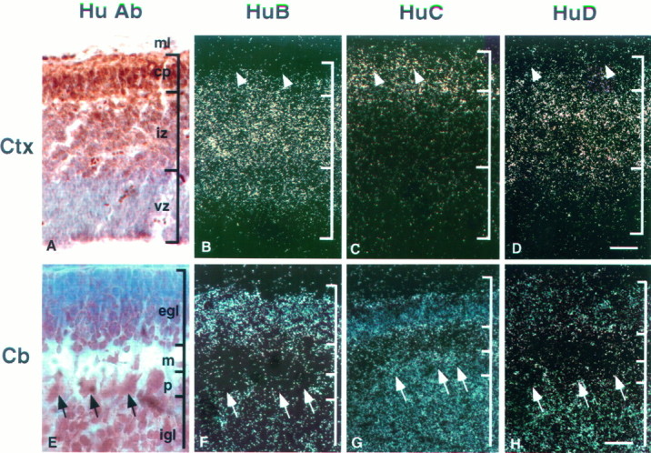
Fig. 7.

Expression patterns of Hu mRNAs in developing brain. Sagittal sections (11 μm) of E14 mouse cortex (Ctx; A–D) and P9 mouse cerebellum (Cb; E–H) were analyzed by immunohistochemistry (A, E) and in situhybridization (B–D, F–H). Affinity-purified Hu antibody was used for immunohistochemistry (A, E). Serial sections were hybridized with 33P-labeled antisenseHuB (B, F), HuC(C, G), and HuD (D, H) gene-specific 3′ UTR cRNA probes. In the developing cortex, mHuB is expressed in some cells of the ventricular zone and cells of the intermediate zone;mHuB diminishes in the cortical plate, with no expression evident in the outermost differentiated neurons (arrowheads). mHuC is detected only in the cortical plate, including the differentiated neurons (arrowheads). mHuD expression is intense in the intermediate zone, diminishes in the cortical plate, and is very weak or absent in the differentiated neurons (arrowheads). In developing cerebellum,mHuB is expressed primarily in the external granule cell layer, whereas the expression of mHuC andmHuD is distributed widely. Purkinje cells (arrows) express only mHuC. Sections were counterstained with cresyl violet. ml, Marginal layer;cp, cortical plate; iz, intermediate zone; vz, ventricular zone; egl, external germinal cell layer; m, molecular layer;p, Purkinje cell layer; igl, internal granule cell layer. Scale bars: 60 μm in A–D; 15 μm in E–H.
