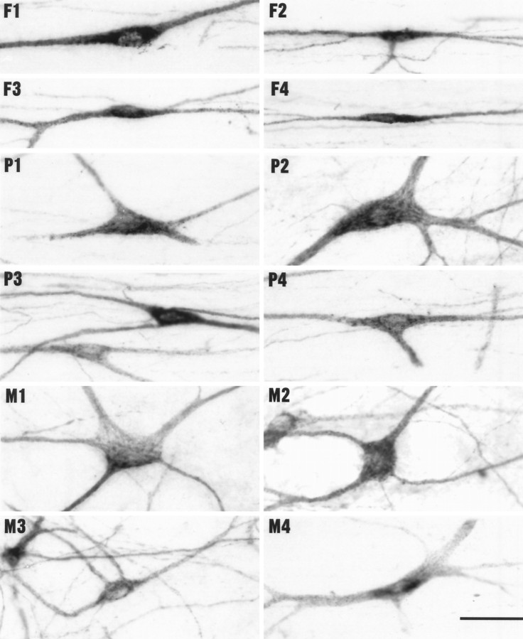
Fig. 4.
Photomicrographs showing varied examples of the three major types of labeled lamina I STT cells in 50 μm horizontal sections (medial up, caudal left).F, Fusiform cells with spindle-shaped somata and two main dendrites; P, pyramidal cells with triangular somata and three main dendrites; M, multipolar cells with polygonal somata and several radiating dendrites. Scale bar, 50 μm.

