
Fig. 1.
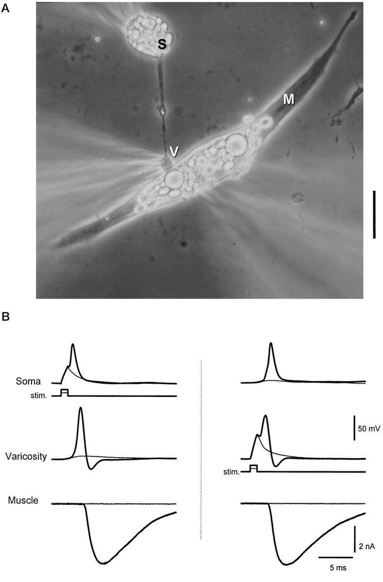
Triple patch-clamp recording from a synaptically coupled cell pair in culture. A, Phase-contrast photomicrograph of a representative preparation. Electrodes for patch recordings are placed on the neuronal soma (S), presynaptic neuronal varicosity (V), and postsynaptic muscle cell (M). Recordings were made in current clamp in the neuron at both locations and in voltage clamp in the muscle. Scale bar, 20 μm. B,Left, A subthreshold depolarization of the neuronal soma (stim.) evoked passive responses in the soma and varicosity and failed to evoke transmitter release (thin traces). Suprathreshold current injection evoked a somal action potential that propagated to the presynaptic varicosity and resulted in the release of transmitter, detected as an endplate current in the muscle cell (thick traces). Right, Direct stimulation of the presynaptic varicosity with a current step evoked an action potential that caused the release of transmitter (thick traces). The locally generated action potential back-propagated to the soma. Subthreshold depolarization of the varicosity did not evoke transmitter release (thin traces). Internal solution A was used for the soma and muscle recording; internal solution B was used for the varicosity recording. Resting potentials were the following: Soma, −65 mV;Varicosity, −69 mV. Similar results were seen in four other preparations.