Fig. 5.
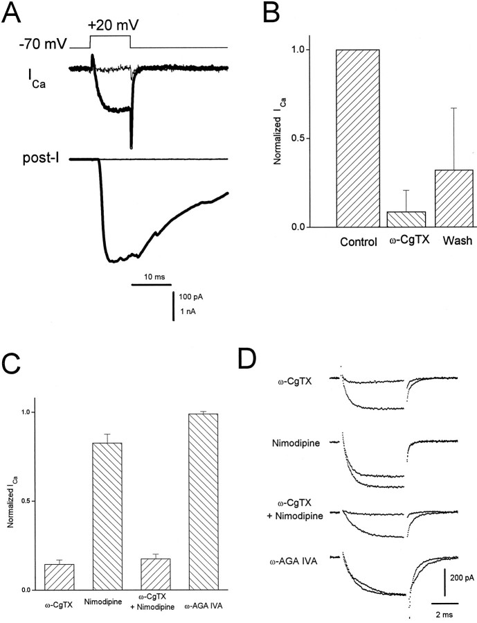
Pharmacology of presynaptic Ca currents and transmitter release. A, Thick traces, A depolarizing step of voltage evoked an inward Ca current (ICa) correlated with a large EPSC (post-I). After application of 1 μm ω-conotoxin GVIA (ω-CgTX), all transmitter release was eliminated, as was most of the Ca current (thin traces). Internal solution C was used in the presynaptic pipette. The external solution was NFR containing 300 nm TTX and 1 mm DAP. B, Bar graph showing the degree and reversibility of block of presynaptic Ca current by 1 μm ω-CgTX (n = 6). In all cases, block of transmitter release was complete, with little or no recovery after wash. C, Bar chart indicating the percentage of varicosity Ca current remaining after application of 2 μm ω-CgTX (n = 8), 2 μm nimodipine (n = 3), ω-CgTX plus nimodipine (n = 4), and 500 nm ω-AGA IVA (n = 5). In these studies, the Ca current was isolated using a combination of 1 μm TTX, 5 mm DAP, and 20 mm TEA in a 10 mmCaCl2 bath solution and using internal solution of the following composition (in mm): 68 CsMeSO4, 50 CsCl, 8 MgCl2, 10 HEPES, pH 7.4. Currents were evoked by steps to +20 mV from a holding potential of −80 mV. D, Current traces (average of four) associated with treatments indicated by the bars in C, taken from representative experiments, showing currents before and after drug addition.

