
Fig. 1.
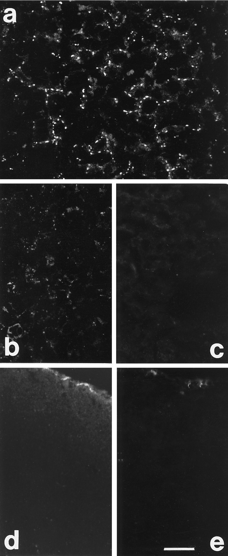
Cx 26 immunoreactivity in liver and brain.a, Labeling localized between hepatic cells in liver sections. b, c, Reduced labeling and absence of staining after treatment of sections with Cx 26 antibody preadsorbed with 20 (b) and 80 μg/ml (c) of peptide, respectively. d, e, Sections through the visual cortex of P7 rats treated with Cx 26 antibody preadsorbed with 20 (d) and 80 μg/ml (e) of peptide, respectively. Note the meningeal labeling (top of section); staining is much reduced in d and completely blocked in e. Scale bar, 60 μm.