
Fig. 2.
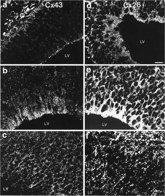
Cxs 26 and 43 immunolabeling in the dorsomedial telencephalic wall of E12–E16 rat brains. a–c, Examples of images taken from sections of E12 (a), E14 (b), and E16 (c) brains labeled for Cx 43. Labeling at the pial surface (P) of the brain ina corresponds to Cx 43 immunoreactivity in meningeal components. d–f, Images from sections of brains of corresponding ages stained for Cx 26. LV, lateral ventricle. Scale bar, 32 μm.