
Fig. 4.
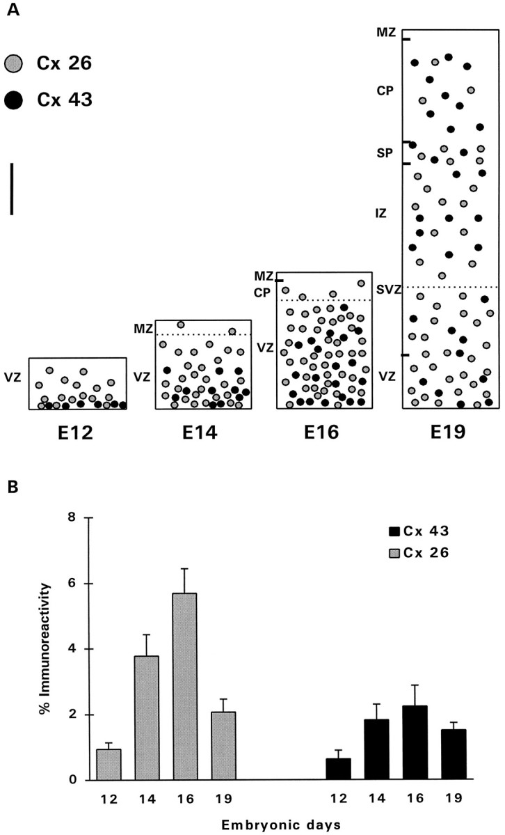
Expression of Cxs in the developing dorsal telencephalic wall of rat embryonic brains. A, Schematic representation of the pattern of distribution of Cx 26 and 43 immunoreactivities at various stages of corticogenesis. At E12, Cx 26 was expressed throughout the neuroepithelium, whereas Cx 43 was localized predominantly between cells bordering the ventricle. At E14–E16, both Cxs showed increased expression throughout the telencephalic wall. At E19, Cx 26 was more concentrated in the proliferative zones (VZ and SVZ), whereas Cx 43 showed a more homogeneous expression through the thickness of the expanding telencephalic wall. VZ, Ventricular zone;MZ, marginal zone; CP, cortical plate;SVZ, subventricular zone; IZ, intermediate zone; SP, subplate. Measurements of immunoreactivity were performed in the VZ at E12–E16, and in both the VZ and SVZ at E19;dotted lines indicate the upper limits of the areas measured. Scale bar, 80 μm. b, Levels of immunoreactivity (%) of Cxs 26 and 43 measured in the proliferative zones of 12 embryonic brains at each age. The measured levels were corrected, taking into account the radial expansion of the developing cortex. Error bars represent SEM.