Fig. 8.
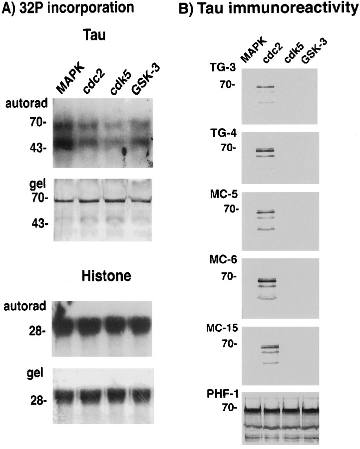
Preferential creation of the TG/MC phospho-epitopes in recombinant tau by cdc2/cyclin B. A,32P incorporation. Histone H1 and tau were incubated with the indicated proline-directed kinases. Bottom panels(i.e., gel) for each substrate show Coomassie blue staining of the protein in the gel, and the top panels (i.e., autorad) show the corresponding autoradiograms. Equivalent amounts of32P were incorporated into H1 after incubation with the indicated kinases. This was also the case with tau as substrate, except that higher amounts of 32P incorporation were observed with MAPK. The positions of molecular weight (MW) markers in kilodaltons are shown on the left of each panel. B, Tau immunoreactivity. Replicate panels of tau protein phosphorylated with MAPK, cdc2/cyclin B, cdk5, or GSK-3, respectively, were stained with the TG/MC and PHF-1 antibodies as indicated on the leftof each panel. Whereas all the kinases produced the PHF-1 phospho-epitope in tau, only cdc2/cyclin B produced the mitotic TG/MC epitopes. The position of the 70 kDa MW marker is shown on theleft of each panel.

