Fig. 2.
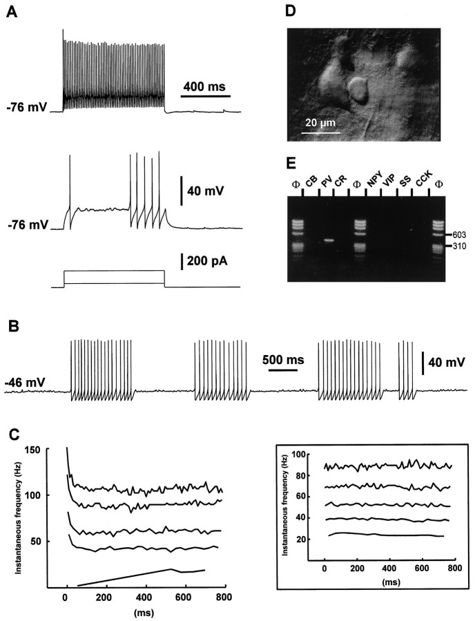
Electrophysiological and biochemical characterization of a layer V FS cell. A, Current-clamp recording during injection of depolarizing current pulses. Membrane potential was adjusted to −76 mV by continuous current injection, as indicated on the left of each recording. In response to a near-threshold current pulse (50 pA; bottom trace), this FS cell emitted a single fast action potential with a large AHP followed by a silent period and a late discharge of action potentials. Note the membrane potential oscillations during the silent period. Application of a larger depolarizing current (200 pA; top trace) induced a continuous discharge at high frequency.B, Continuous near-threshold depolarizing current (60 pA) evoked clusters of nonaccommodating discharges of fast action potentials. Note the membrane potential oscillations between these clusters. C, Left panel, Analysis of the instantaneous firing frequency during the action potentials discharges evoked in the same FS cell by current pulses of increasing intensities (50–250 pA; bottom to top traces). Note that after a fast early accommodation, discharges rapidly reached a steady-state frequency. Inset, Analysis of the instantaneous firing frequency during the action potentials discharges evoked in another FS cell by current pulses of increasing intensities (50, 100, 150, 200, and 350 pA; bottom to top traces). Note the lack of early accommodation of the discharge frequency at each stimulation intensity. D, IR videomicroscopy picture of the FS cell. The FS cell is located at the center and has a small round soma (diameter ∼10 μm). Note the large layer V pyramidal cell immediately on the left. Pial surface isupward. E, Agarose gel analysis of the RT-mPCR products of the same FS cell. The only PCR-generated fragment was that of PV.

