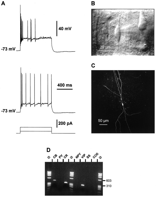
Fig. 5.
Physiological, morphological, and RT-mPCR analysis of a layer V IS cell. A, Current-clamp recording obtained in response to depolarizing current pulses (50 and 150 pA). Note the initial burst of action potentials, the irregular firing frequency of the following spikes, and the membrane potential oscillations between each action potential. B, IR videomicroscopy of this cell typically presenting a vertically oriented fusiform soma (20 μm long). Two main dendrites extended in opposite directions from the soma, one toward the superficial layers and the other toward the white matter. C, Confocal image of the cell labeled by intracellular biocytin injection. This IS bipolar cell displayed two main narrow dendritic arborizations. The ascending dendrites (top) extended up to layers II–III; the descending dendrites extended to layer VI. D, Agarose gel analysis of the second PCR products. Three fragments corresponding to CB, CR, and VIP with a size of 432, 309, and 287 bp, respectively, were amplified.

